Corneal Endothelial Dystrophy
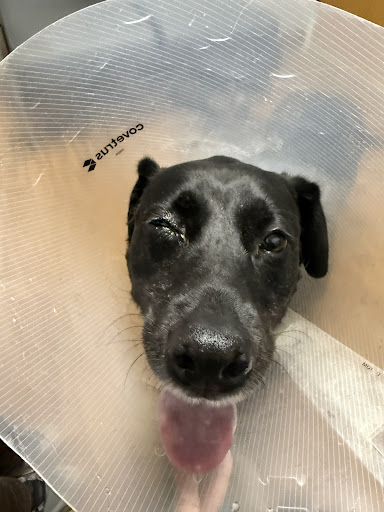
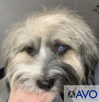
 The hallmark sign of corneal endothelial dystrophy in dogs is eyes with a blue or foggy appearance. As the disease progresses, patients experience ocular discomfort and pain. Your pet may also avoid bright light or show signs of visual discomfort when outdoors.
While corneal endothelial dystrophy is a progressive eye disease in dogs, treatment options are available from medications to advanced surgical procedures.
At Armour Veterinary Ophthalmology, we offer the latest approach to restore vision and comfort for dogs with corneal endothelial dystrophy, called DSEK.
Whether you’ve been referred to us by your primary veterinarian or just want a second opinion regarding this disease, our team is here to help!
What is Corneal Endothelial Dystrophy?
Corneal endothelial Dystrophy or Endothelial Degeneration is a disease of the internal layer of the cornea. In a normal cornea, endothelial cells work as a pump to remove fluid from the cornea. This maintains the cornea’s clear appearance.
When endothelial dystrophy is present, endothelial cells are gradually lost. This means fluid is no longer removed from the cornea. This results in corneal edema (swelling), cloudiness of the cornea, and decreased or “blurred” vision.
With chronic (long-term) edema, the fluid accumulates, forming pockets, known as bullae. These bullae can rupture through the outer layer of the cornea (the epithelium), forming a corneal ulcer. These ulcers are very painful, may be recurrent, and often non-healing.
There are limited medical and surgical options available for treatment of endothelial dystrophy. Medical therapy consists of using topical drops such as a topical anti-inflammatory medication.
In past years, the treatment of choice was a thermokeratoplasty as an attempt to prevent ulcer formation. This involves using a small cautery unit to heat small spots of the affected cornea. These areas will then form scars, which prevent the spread of the fluid-filled bullae.
Another surgical option is to place a conjunctival flap or gunderson flap. This consists of making a flap of tissue from the conjunctiva (pink tissue surrounding the eye) and suturing it over the affected areas of the cornea. This procedure relieves the discomfort caused by corneal ulceration, however it will leave a scar on the cornea, and does not address the main cause of the corneal changes.
The last surgical option is a corneal transplant (similar to the procedure done in humans). Corneal endothelial dystrophy is similar to Fuch’s endothelial corneal dystrophy (FECD).
Before Treatment 
After Treatment

Before Treatment After Treatment


Endothelial keratoplasties (EKs), in particular Descemet’s Stripping Endothelial Keratoplasty (DSEK) is a novel surgical technique for corneal endothelial dystrophy in veterinary ophthalmology.
Here at Armour Veterinary Ophthalmology, we have been pioneering DSEK as a procedure to restore vision and comfort for dogs with corneal endothelial dystrophy.
If this is a procedure that you would like to consider, we would love to set up an in-depth phone conversation with you.
Located inside the Friendship Hospital for Animals in Washington D.C., we serve patients throughout the greater Washington D.C. area including Maryland and northern Virginia.
If your pet has been diagnosed with corneal endothelial dystrophy, our team can help!
Our practice is conveniently located in the Friendship Hospital for Animals in Washington D.C. We serve patients throughout Maryland and northern Virginia.